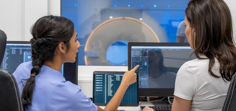
What is an MRI?
MRI or magnetic resonance imaging uses a strong magnetic field and radio waves to create a detailed pictures of areas inside the body. The equipment creates images of the breast or other organs from every angle, including side to side and top to bottom. A radiologist interprets the images in making a diagnosis.
What are MRIs used for?
- Breast imaging: provide a clearer view of a mass or suspicious area for accurate diagnosis
- Prenatal fetal imaging: further evaluation of fetal abnormalities detected on ultrasound
- Premature or ill infant imaging: evaluation of brain and spine abnormalities which are often better demonstrated with MRI as compared to CT scan or ultrasound
- Adult female pelvis imaging: evaluation of patients with ovarian cancer, uterine fibroids and conditions that cause infertility
Closed vs. Open MRI
While entering an enclosed space can be intimidating, closed MRI equipment contains a more powerful magnet than "open" MRI systems, meaning the images it produces are more detailed, more precise and feature greater resolution.
The closed MRI machine at Woman's combines the shortest possible tube length with a high-strength scanner to reduce patient concerns without sacrificing image quality.
The shorter tube length means you may be able to tolerate the procedure a lot better than in a regular MRI machine that is longer in length. Your body does not have to go so far into the tube, so you get the higher resolution of the closed MRI with the less claustrophobic aspects of the open MRI.
MRI and Breast Cancer Detection
Your doctor may recommend a MRI or a MRI-guided breast biopsy if mammogram and ultrasound do not provide a clear enough view of a mass or suspicious area for an accurate diagnosis.
Who may need a breast MRI?
You doctor will determine if breast MRI is right for you based on your health history.
Your doctor may recommend it if you:
- Have a known diagnosis of breast cancer
- Carry the genes BRCA1 or BRCA2, which put you at increased risk for breast cancer Have breast implants
- Have scars from previous surgeries
What you can expect?
MRI’s are not painful or physically uncomfortable.
You will lie on your stomach on the scanning table to allow your breasts to drop into a hollow depression. Inside the depression is a coil that detects a magnetic signal. The table then moves inside a large tube that contains the magnet. The patient is given an injection of contrast agent to improve the clarity of the images of the tissue or tumor.
Imaging sessions typically take about one hour.